



By Dr. Dion Paridaens (ophthalmologist)
Dear Readers,
Last year, the two-volume European (ESOPRS) textbook on Eyelid, Lacrimal, and Orbital Surgery was published. I contributed to this work as an editor and author, alongside colleagues from Europe and other parts of the world.
For the OOG in OOG Association, with which I have been involved as a medical advisor for many years, I have rewritten some key topics from this book in easy-to-understand language. I will share these summaries with the readers of the association’s magazine.
This summary is about enucleation (the surgical removal of an eye). It provides an overview of the key points, such as:
- when this procedure is necessary,
- how the preparation and the surgery itself proceed,
- and what kind of post-operative care is important.
The full text in the book is more detailed and intended for specialists. We hope you find this summary helpful. More summaries of other chapters from the European textbook will follow shortly.
Sincerely,
Dion Paridaens
Reasons for removing an eye:
- There is a tumor in the eye that cannot be treated with medication.
- My eye is blurry and very painful, and I need to get it checked out to find out the cause.
- The eye has been severely damaged in an accident, which increases the risk of a dangerous inflammatory reaction in the other eye.
When is enucleation not recommended?
- If the eye cancer has already spread beyond the eye. In that case, it may be necessary to remove not only the eye but also the surrounding tissue (orbital exenteration).
About the Surgery
- Restoring volume: Because the eye is removed, an empty space is created in the eye socket. For this reason, an implant (an artificial ball) is almost always placed to preserve the shape of the face.
- Attaching the eye muscles: The eye muscles are attached to the implant. This helps the prosthetic eye move later on and prevents the implant from shifting.
- Preventing complications: Close attention must be paid to bleeding to prevent problems such as a blood clot (hematoma) or displacement of the implant. The wound is carefully closed.
- Operating on the correct eye: Because this is a major surgery, checks are performed multiple times to ensure that the correct eye is being removed.
Preparing for Surgery
- Medical examination: The doctor examines the eye and performs scans (such as an MRI or ultrasound) to determine whether the tumor has spread.
- Assess overall health: For example, if the patient has diabetes or has previously undergone radiation therapy, this may affect the healing process.
- Psychological counseling: Losing an eye can be emotionally difficult. The doctor therefore explains in detail what the patient and their loved ones can expect and what options are available for a prosthesis.
How is the surgery performed?
- Preparation: The appropriate eye is examined and marked. The patient is positioned correctly.
- Detachment of the eye tissues: The outer layers of the eye are carefully detached. The eye muscles are also prepared for later attachment to the implant.
- Removal of the eye: The eye is cut free and removed. If there is a tumor, an additional section of the optic nerve is removed to investigate whether the cancer has spread.
- Implant Placement: After the eye is removed, an implant is placed. This may be a small sphere made of a special material that is well tolerated by the body.
- Closing the Incision: The eye muscles are attached to the implant, and the incision is carefully closed. A temporary prosthesis is inserted to maintain the shape of the eye socket.
After the surgery
- Immediately after enucleation: either a temporary ocular prosthesis or a temporary conformer is inserted. Both serve to shape the space for the permanent ocular prosthesis and maintain the depth of the conjunctival folds, which are important for a proper fit.
- Immediately after the procedure: The patient is given pain medication and antibiotics to prevent infection. A compression bandage is applied to reduce swelling. The patient is usually able to go home the next day.
- Recovery and Aftercare: The temporary prosthesis remains in the eye socket for several weeks. The patient receives instructions on how to care for the wound and how to handle the other eye with care. After 6 to 8 weeks, a permanent ocular prosthesis is fitted.
Which implant should be chosen?
- Size: The implant must be large enough to replace the volume of the removed eye. For adults, an implant measuring 20–22 mm is usually used.
- Material:
- Non-porous materials (such as silicone or PMMA) are less prone to inflammation.
- Porous materials (such as hydroxyapatite) promote better integration of the body’s own tissue, but can sometimes become inflamed more quickly.
- Additional options:
- Wrapping: Sometimes a thin layer of donor tissue is placed around the implant to help it adhere better.
- Movable prosthesis (pegging): In some cases, a small pin is used to make the artificial eye prosthesis more mobile. However, this procedure is performed less frequently due to a higher risk of complications.
Conclusion
- Enucleation is a surgical procedure in which the eye is completely removed, often due to cancer or a severely damaged eye.
- An implant is placed to restore volume in the eye socket and preserve the shape of the face.
- Good follow-up care and support are important to help the patient adjust to an artificial eye.
- The choice of implant depends on the situation and the surgeon’s preference, with the safest option carrying the fewest risks being selected.
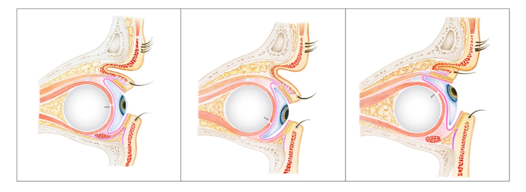
Figure 1: Movement of the implant and the ocular prosthesis after enucleation
When a person looks straight ahead, the ocular prosthesis is centered and the pupil is clearly visible (left). Because the eye socket is not deep enough and there is no direct connection between the prosthesis and the implant, the prosthesis is less able to move with the eye. This is particularly noticeable when looking down (center) and looking up (right).
(from: Ntountas, I., Tsirouki, T., Klett, A., Sigurdsson, H., Paridaens, D. (2024). Enucleation. In: Quaranta Leoni, F.M., Verity, D.H., Paridaens, D. (eds) Oculoplastic, Lacrimal, and Orbital Surgery. Springer, Cham. https://doi.org/10.1007/978-3-031-39638-0_1
